
Neurophysiology

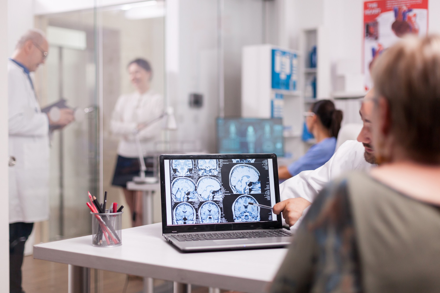
Neurophysiology is the study of how the brain, spinal cord, nerves, and muscles function and communicate with one another. Neurophysiological testing, including electromyography (EMG), electroencephalography (EEG), and nerve conduction studies, helps healthcare professionals evaluate neurological symptoms, identify abnormalities in nerve and muscle activity, and assess brain function. These tests support the diagnosis and management of conditions such as epilepsy, nerve injuries, muscle disorders, and peripheral neuropathy, helping guide appropriate treatment and ongoing patient care.

Neurophysiology Diseases Q&A

What Is an EMG?

An electromyography (EMG) test measures the electrical activity of muscles and helps evaluate the health of the nerves that control them. It is commonly used to investigate symptoms such as muscle weakness, numbness, tingling, cramps, and unexplained muscle pain.
During the test, a healthcare professional may insert a thin needle electrode into selected muscles to record their electrical activity at rest and during contraction. The results can help identify nerve or muscle disorders and assist with diagnosing conditions that affect the connection between nerves and muscles.

Types of EMG Tests

Electromyography (EMG) tests evaluate muscle electrical activity and help identify problems affecting nerves and muscles.

  • Needle EMG: Uses a thin needle electrode inserted into selected muscles to measure electrical activity at rest and during contraction.
  • Surface EMG: Uses electrodes placed on the skin to record muscle activity without inserting needles.
  • Nerve Conduction Study (NCS): Often performed alongside EMG to measure how quickly and effectively electrical signals travel through peripheral nerves.
  • EMG with Nerve Conduction Studies: Combines muscle testing and nerve signal measurements to help evaluate conditions such as carpal tunnel syndrome, peripheral neuropathy, and nerve injuries.

What Is an EEG?

An electroencephalogram (EEG) is a noninvasive test that records the brain’s electrical activity. It is often used to help evaluate seizures, epilepsy, unusual episodes of lost awareness, and other conditions that may affect brain function.

During the test, small electrodes are attached to the scalp using sensors or a special cap. These electrodes record electrical signals from the brain while the patient rests, and sometimes during activities such as opening and closing the eyes, deep breathing, or viewing flashing lights. EEG results help healthcare professionals identify unusual brain activity and support diagnosis and treatment planning.

Types of EEG Tests

Electroencephalogram (EEG) tests record electrical activity in the brain and help evaluate seizures, epilepsy, and other neurological conditions.

  • Routine EEG: A standard recording that usually lasts around 20–60 minutes and may include eye-opening, deep-breathing, or flashing-light activities.
  • Sleep EEG: Records brain activity during sleep or drowsiness to identify abnormalities that may not appear while awake.
  • Sleep-Deprived EEG: Performed after reduced sleep to increase the chance of detecting certain abnormal brain patterns.
  • Ambulatory EEG: Uses a portable recording device to monitor brain activity during everyday activities, often for 24–72 hours or longer.
  • Video EEG: Combines EEG recording with video to help doctors compare brain activity with a patient’s movements and behavior.
  • Continuous EEG (cEEG): Provides prolonged brain activity monitoring, commonly in hospitalized patients who need ongoing neurological assessment.

What Is an EMU?

An Epilepsy Monitoring Unit (EMU) is a specialized hospital unit where patients undergo continuous monitoring to evaluate seizures, suspected epilepsy, and unexplained episodes of altered awareness or unusual movements. It allows specialists to study brain activity and physical symptoms together to better understand what is happening during these episodes.

During an EMU stay, the patient is connected to EEG electrodes that continuously record brain activity, while video monitoring captures physical movements and behavior. This combined video-EEG monitoring helps doctors determine whether episodes are epileptic seizures, identify seizure types, understand where seizures may begin in the brain, and plan appropriate treatment. Hospital stays often last several days, depending on the information needed. Any changes to seizure medication are managed by the medical team under supervision.

Types of EMU Monitoring

An Epilepsy Monitoring Unit (EMU) is a specialized hospital setting rather than a single test. It provides continuous monitoring to investigate suspected seizures and help guide epilepsy treatment.

  • Inpatient Video EEG Monitoring: Records brain activity and synchronized video while the patient stays in the hospital.
  • Long-Term EEG Monitoring: Records brain activity over an extended period to capture and evaluate seizure events.
  • Seizure Localization Monitoring: Helps specialists identify the area of the brain where seizures may begin.
  • Medication Adjustment Monitoring: Allows the medical team to assess seizure activity while making supervised changes to antiseizure medications when clinically appropriate.
  • Pre-Surgical Epilepsy Monitoring: Helps determine whether epilepsy surgery may be appropriate by gathering information about seizure onset and patterns.
  • Event Characterization Monitoring: Helps distinguish epileptic seizures from other episodes, such as fainting or certain nonepileptic events.

Accessibility Toolbar